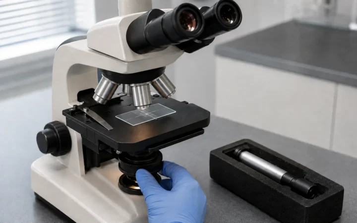

Best Microscopes for Pathology Labs in 2026 connects microscope specifications to real laboratory workflow, emphasizing optical performance, calibration, documentation, serviceability and the needs of the actual method.
Quick Verdict
- Best for: labs connecting instrument specifications to diagnostic or research workflow.
- Focus on: optics, illumination, camera/digital needs, calibration, service and documentation.
- Watch for: premium features add little value when they do not support the method or quality system.
Table of contents
Introduction
Pathology is the cornerstone of modern medicine. By examining tissue sections under a microscope, pathologists diagnose cancers, assess surgical margins and monitor disease progression. Choosing the right microscope for a pathology lab can dramatically influence diagnostic speed, workflow efficiency and even the health of laboratory professionals who spend hours at the eyepiece. Yet selecting a microscope is not straightforward—manufacturers offer dozens of models, each with different optics, illumination and ergonomic features. In this guide we’ll demystify the process by examining critical features, highlighting the top microscopes available in 2026 and explaining how digital pathology and automation are reshaping the field. We’ll also link to related resources on FrediTech and answer common reader questions.

Why microscope selection matters in pathology
Tissue slides must be examined with crystal‑clear, color‑faithful images to discern subtle histological features such as mitotic figures, inflammatory infiltrates or abnormal cellular architecture. According to ZEISS, histopathologists rely on microscopes that provide “the highest color fidelity in brightfield” and the optical quality of the microscope and attached camera are critical for accurate screeningzeiss.com. Leica stresses that pathologists spend long hours at their microscopes, so ergonomics and automation of routine functions are essential for efficiency and to prevent injuries. Rapid workflows also depend on integrated software: Leica’s DM4 B and DM6 B microscopes use Synapse technology to synchronize components, boosting productivity up to two times compared with traditional upright microscopesleica-microsystems.com. Nikon highlights that its upright microscopes combine high optical performance, ergonomics and cameras capable of high‑resolution, true‑color imaging that can be shared remotelymicroscope.healthcare.nikon.com. In short, selecting a microscope isn’t just about magnification; it’s about how the instrument supports a pathologist’s workflow and well‑being.
Key features to consider
Choosing a pathology microscope involves balancing optical performance, ergonomics, automation, digital connectivity and budget. The following sections break down the core features you should evaluate.
Optical quality and contrast methods
Accurate diagnosis requires microscopes with high numerical aperture objectives that render crisp images and preserve color fidelity. ZEISS points out that . High‑quality optics are especially important for techniques such as immunohistochemistry (IHC) and fluorescence. Many microscopes provide multiple contrast methods—brightfield, phase contrast, polarized light, fluorescence and differential interference contrast (DIC). For example, Olympus’ BX53 series supports multi‑color fluorescence imaging and phase contrast, while its BX53M offers advanced contrast and imaging options with modular materials‑science featuresevidentscientific.com.
Long working distances and wide fields of view also enhance usability. Evident’s upright microscopes offer an FN26.5 ocular with an industry‑leading field of view for immersive observation. Wide fields reduce the number of stage movements needed to scan a slide and make it easier to contextualize small lesions within surrounding tissue.
Illumination systems
Ergonomics and user comfort
Stage and focus controls should be symmetrical to prevent twisting and reaching. Leica’s ergonomics guide recommends symmetrical stage drive and focus knobs so users maintain level shoulders and a straight spine. The ability to switch stage controls from right‑handed to left‑handed operation (or vice versa) is also valuable when multiple technicians share a microscope.leica-microsystems.com
Automation and digital documentation
Digital connectivity is a growing requirement. Nikon notes that its upright microscopes provide clear, high‑resolution images and superior color reproduction across a wide field of view, and . Remote sharing facilitates telepathology and collaborative diagnosis. Zeiss’s smart microscopy can integrate a barcode reader to assign scaling information to labelled slides, streamlining data management.
Budget and total cost of ownership
While high‑end microscopes can cost tens of thousands of dollars, total cost of ownership includes service, warranty and software licensing. LED illumination reduces lamp replacement costs, and energy‑saving features like Zeiss’s Eco‑mode lower electricity bills. Labs should also consider the cost of digital cameras, slide scanners and software subscriptions when budgeting for a new system.
Top microscopes for pathology labs
Based on the features above and current market offerings, the following microscopes stand out for pathology in 2026. Each section summarizes the key strengths of the model and provides real‑world context.
Leica DM4 B & DM6 B – Customizable workhorses for tissue and plant pathology
Real‑world example: A teaching hospital pathology lab in Johannesburg upgraded to the DM6 B in 2025 to handle high case volumes. Technologists appreciated the automated stage and Synapse‑driven workflow, which reduced slide handling time. The built‑in LAS X Navigator allowed them to generate mosaic images of large specimens quickly, and the AI analysis module flagged suspicious areas for pathologist review. Within months, the lab’s turnaround time for biopsy reports decreased by 20 %, and ergonomics improved because staff no longer had to contort to operate manual knobs.
Olympus BX™ series – Modular design and True‑Color imaging
Real‑world example: A regional hospital in Ghana adopted the BX46 for routine histology. Technicians quickly noticed the difference: the tilting telescopic tube allowed each user to adjust the eyepiece height, reducing neck strain. Combined with the ultra‑low stage, the microscope allowed comfortable operation for technologists of various heights. When the lab later added immunofluorescence testing, they upgraded the BX46 with a fluorescence module and camera—demonstrating the value of modularity.
Nikon upright microscopes – Reliable optics and remote collaboration

Nikon’s clinical research microscopes deliver high optical performance and are optimized for repeated use. Their design emphasizes usability and comfort, and Nikon color cameras provide clear, high‑resolution images with . Importantly, Nikon highlights that images can be shared remotely in real time, facilitating virtual consultations. Nikon’s line includes the ECLIPSE Ci series for routine use and the more advanced ECLIPSE Ni series with motorized options and digital integration. While Nikon does not list specific models on its “clinical research” page, the company is renowned for mechanical reliability and durable build quality, making it a strong choice for labs that need robust instruments with minimal downtime.
Real‑world example: During the COVID‑19 pandemic, a private pathology lab in Accra upgraded to Nikon Ci‑L microscopes with DS‑Fi3 cameras. The combination allowed pathologists to share live images with colleagues working remotely, enabling consultations on complex cases. The lab integrated the system with teleconferencing software, which improved diagnostic confidence and allowed for real‑time teaching of junior residents.
Zeiss Axiolab 5 – Smart microscopy for efficient documentation

Real‑world example: A digital pathology startup in Lagos implemented Axiolab 5 microscopes to create a hybrid workflow between manual screening and slide scanning. The smart documentation features helped them quickly generate digital images for remote AI analysis. The built‑in Eco‑mode fit their sustainability goals, while ergonomic controls minimized operator fatigue during long scanning sessions.
Zeiss Axioscope 5 – Multichannel fluorescence made simple
Zeiss’s Axioscope 5 is a “smart laboratory microscope” designed for histology and fluorescence. The company states that this microscope enables acquisition of fluorescent images in up to four different channels and includes an adjustable ergotube to match users’ natural posture. Combining ergonomic features with automation, the Axioscope 5 is well‑suited for labs that need fluorescence capability without the complexity of confocal microscopes.
Emerging player: Fein Optic RB40 4K Pathology Lab Microscope
While not from a major manufacturer, the Fein Optic RB40 4K microscope is noteworthy. Microscope World describes it as being equipped with a 4K HD scientific‑grade camera that streams live video and paired with a 40 × Plan Semi‑Apochromat objective ideal for pathology workmicroscopeworld.com. Although primarily a niche product, the inclusion of a 4 K camera and dedicated monitor could appeal to labs seeking high‑resolution digital viewing without external cameras. Users should evaluate vendor support and service availability before purchasing.
Step‑by‑step guide to choosing a pathology microscope
Selecting a microscope involves systematic evaluation. Follow this step‑by‑step process to ensure the chosen system aligns with your lab’s clinical needs, workflow and budget.
Step 1: Define your primary applications
List the types of specimens you handle (e.g., hematoxylin and eosin staining, immunohistochemistry, frozen sections, plant tissues). Identify whether fluorescence, phase contrast or polarized light will be used. If digital pathology or telepathology is a goal, include digital imaging requirements (camera resolution, slide scanning).
Step 2: Determine optical and illumination requirements
Choose microscopes with objectives appropriate for your magnification needs (commonly 4 ×, 10 ×, 20 × and 40 ×; oil‑immersion 100 × for fine detail). Ensure the microscope offers high numerical aperture lenses and a wide field of view. LED illumination is preferred for consistent brightness and energy efficiency; ensure the system provides true‑color rendering and supports various contrast techniques.
Step 3: Prioritize ergonomics
Evaluate whether the microscope has adjustable eyepieces (tilting and telescopic tubes), height‑adjustable stages and symmetrical controls. According to Leica, an ergonomic design customized to the user’s body size promotes good posture and minimizes strain. Consider models with low‑profile stages or tilting tubes (e.g., Olympus BX46 or Zeiss Axiolab 5).
Step 4: Assess automation and software integration
Automated focusing, motorized nosepieces and slide scanning can significantly improve throughput. Leica’s Synapse technology synchronizes components and doubles productivity. Zeiss’s smart microscopy automatically adjusts camera settings and adds scale bars. Determine whether you need integrated AI analysis (e.g., AIVIA) or digital slide scanning capabilities.
Step 5: Evaluate digital imaging and connectivity
Digital cameras should provide high resolution (ideally 5 MP or more), true color reproduction and minimal noise. Nikon notes that its microscopes deliver images that can be shared remotely in real time, enabling teleconsultation. Check for compatibility with your laboratory information system (LIS) and ensure that image capture is simple and reliable.
Step 6: Consider future scalability and service
Choose a microscope that can be upgraded with new modules (e.g., fluorescence illuminators, motorized stages) and ensure that parts and service are available locally. Evaluate warranty terms, training support and whether the vendor provides software updates.
How digital pathology is transforming microscopes
Digital pathology involves converting glass slides into digital images for analysis, sharing and storage. This shift has accelerated since the COVID‑19 pandemic. According to a 2025 market report, the global digital pathology market was valued at USD 1.15 billion in 2024 and is projected to grow from USD 1.30 billion in 2025 to USD 3.86 billion by 2032, a compound annual growth rate (CAGR) of 16.9 %. The report notes that digital tools increased productivity by 15 % compared with traditional workflows and that adoption surged during the COVID‑19 pandemic due to remote consultation needs. Whole slide scanners and digital microscopes capture high‑resolution images that can be annotated and processed with AI algorithms. This reduces manual slide handling, allows remote review and creates large datasets for machine learning. Leading microscope manufacturers now offer integrated cameras, slide scanners and AI software (e.g., Leica’s AIVIA, Zeiss’s smart microscopy). Labs adopting digital pathology should consider microscopes with seamless digital workflows and standardized data formats.
Real‑world case study: Implementation of digital microscopy in a Ghanaian hospital
In 2025, a tertiary hospital in Kumasi launched a digital pathology pilot. The laboratory replaced its 15‑year‑old microscopes with Leica DM6 B units equipped with AIVIA AI analysis. Slides were scanned using an integrated slide scanner, and digital images were stored in the hospital’s LIS. Pathologists accessed slides via tablets and could annotate images during multidisciplinary meetings. The impact was significant:
- Reduced turnaround time: average biopsy report time decreased from 5 days to 3 days.
- Improved collaboration: remote specialists could review slides and discuss findings simultaneously.
- Enhanced training: residents used archived digital slides to study uncommon cases and receive feedback.
- Quality assurance: the system allowed automated quality checks, ensuring focus and color calibration.
However, the transition required training staff, upgrading network infrastructure and ensuring data security. The hospital worked closely with the vendor to customize workflows and integrate AI analysis for detecting mitotic figures. This example underscores that digital pathology is not just about hardware but also about change management and software integration.
Potential challenges and solutions
Challenge 1: High initial cost. Advanced microscopes and digital slide scanners can be expensive. Solution: start with modular systems that allow upgrades, such as Olympus BX43 or Leica DM4 B. Seek government grants or partnerships for digital pathology initiatives.
Challenge 2: Data storage and management. Whole slide images are large. Solution: invest in scalable storage, compress images using efficient formats and implement cloud‑based archival solutions with proper security.
Challenge 3: Training and adoption. Staff may resist new workflows. Solution: provide comprehensive training, highlight ergonomic benefits and productivity gains, and involve users in the selection process. Consider appointing digital pathology champions within the lab.
Challenge 4: Regulatory compliance. In many countries, digital pathology systems require regulatory approval for primary diagnosis. Solution: ensure that your vendor provides systems compliant with local regulations and guidelines.
FrediTech resources
For a deeper dive into digital microscopy, read
- “Complete Guide to Digital Microscopy: Unleashing the Future of Imaging”freditech.com. The article explains how digital microscopes work, outlines components such as objective lenses, sensors and software, and discusses applications from clinical pathology to forensic science. It also explores sampling and quantization, illustrating how image resolution is determined by pixel count and bit depth. This resource complements the current guide by providing foundational knowledge about digital imaging systems. Another recommended post,
- “Advanced Imaging Techniques Transforming Visualization in Medicine, Industry and Beyond,”freditech.com covers emerging modalities like 3D scanning and AI‑driven image analysis.
FAQ
What type of microscope is used for pathology?
How much magnification is needed to view pathology slides?
Are digital microscopes better than traditional optical microscopes?
How important is ergonomics in microscope selection?
What is digital pathology and why is it growing?
Which microscope brand is best for a small clinic?
Can I upgrade my existing microscope to digital?
Conclusion
Selecting a microscope for a pathology lab is a decision that affects diagnostic accuracy, workflow efficiency and the well‑being of lab staff. Key considerations include optical quality, illumination, ergonomics, automation and digital connectivity. Leading manufacturers—Leica, Olympus (Evident), Nikon and Zeiss—offer a range of models that address these needs. Leica’s DM4 B and DM6 B deliver customizable, productivity‑boosting workflows. Olympus’ BX series combines modularity with True Color LED illumination. Nikon microscopes provide reliable optics and seamless remote sharing. Zeiss’s Axiolab 5 exemplifies smart microscopy with one‑button digital documentation and energy‑saving features. Meanwhile, emerging players like Fein Optic RB40 demonstrate the increasing availability of 4K digital imaging for pathology.
Beyond hardware, the future of pathology lies in digital integration. The digital pathology market’s projected CAGR of 16.9 % underscores the rapid shift toward whole slide imaging, AI‑driven analysis and remote collaboration. Laboratories should therefore select microscopes that can evolve with these trends—systems that combine ergonomic excellence, high‑quality optics and smart digital workflows. By doing so, they will not only enhance diagnostic accuracy and efficiency but also protect the health of the professionals whose eyes interpret the microscopic world.
Author
Wiredu Fred – Medical technology analyst, technophile and lead author at FrediTech. Fred holds a Bachelor of Science in Molecular Biology & Biotechnology and has spent over a decade reviewing laboratory equipment and imaging technologies. He’s passionate about helping labs in Africa and beyond adopt modern tools that improve diagnostic accuracy and efficiency..
Bottom Line
Good microscopy decisions connect optics and digital features to the actual method, quality requirements and support environment. Calibration, maintenance and staff competence are part of performance—not afterthoughts.