
Introduction
Microscopy has underpinned scientific discovery for centuries. From Anton van Leeuwenhoek’s handcrafted lenses to the sophisticated electron microscopes of today, being able to see the unseen has propelled biology, medicine and materials science forwardfreditech.com. In 2025 the microscopy market was estimated at USD 13.52 billion and analysts expect it to surpass USD 27.58 billion by 2034 with a compound annual growth rate of 7.39 %precedenceresearch.com. Much of this growth is driven by digital and ergonomic microscopes that simplify observation and documentation.
Olympus’ CX series, launched in 2017, exemplifies this trend. The CX43 and CX33 microscopes were designed “from the user’s perspective,” providing comfortable observation, versatile imaging methods and low running costsolympus-global.com. This review explains what makes these microscopes appealing for laboratories and educational institutions. We will cover their optical performance, ergonomic design, illumination system and practical applications. Along the way you’ll find step‑by‑step explanations, real‑world examples, internal links to helpful FrediTech guides and citations to credible sources. Whether you’re researching your next lab purchase or simply curious about modern microscopes, this comprehensive guide will help you decide whether the CX43 or CX33 suits your needs.

Table of contents
Why digital and ergonomic microscopes matter
Traditional microscopes require prolonged peering into an eyepiece, which can lead to neck strain and fatigue during long sessions. Digital microscopes replace the eyepiece with a high‑resolution camera, sending images to a monitor or computer. This allows multiple people to view specimens simultaneously and enables remote collaborationfreditech.com. The CX43 and CX33 maintain the optical quality of compound microscopes while offering optional camera ports for digital imaging. Their low‑positioned stage and focus controls reduce stress and support long‑term observationsolympus-global.com. Such ergonomic improvements make microscopes more accessible to students and clinicians alike.
Overview of Olympus CX43 and CX33 microscopes
The CX43 and CX33 are part of Olympus’ CX series of upright microscopes. Both models are designed for routine clinical work, educational training and basic research. They share many features—an infinity‑corrected UIS2 optical system, LED illumination and versatile observation methods—but there are important differences to consider.
CX43 key features
- Ergonomic design: The CX43 has a lower stage position (70 mm lower than previous models) to create space under the eyepiece, making it easier to check or swap specimensolympus-global.com. The stage knob and focus knob are positioned lower so they can be manipulated with hands resting on the desk. This design reduces fatigue during long sessions.
- Universal condenser: A universal condenser supports objectives from 2× to 100× and allows brightfield, darkfield and phase‑contrast observations without swapping the condenserolympus-global.com.
- LED illumination: The built‑in LED provides natural color reproduction and has an estimated lifespan of 60 000 hours. Fixed Köhler illumination ensures the right illumination level without adjusting the condensermicro-pro.com.
- Five‑objective nosepiece: A quintuple nosepiece holds up to five objectives, enabling a wide range of magnificationsmicro-pro.com.
- Optional camera port: The CX43 can be fitted with a camera port for digital imaging.
- Weight and dimensions: The microscope weighs approximately 7.3 kg and measures 211 × 376 × 393 mm in its standard configuration.
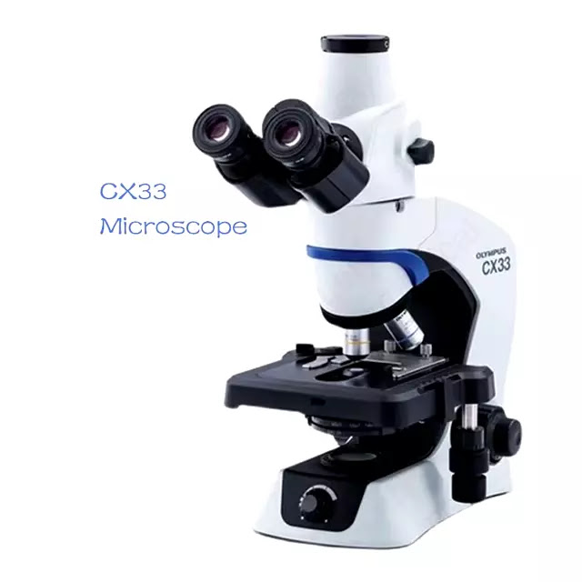
CX33 key features
- Similar ergonomic focus: The CX33 also offers a low‑positioned stage and focus controls for comfortable observation. The controls allow users to set specimens with one hand and adjust focus with the otheroptics-pro.com.
- LED illumination with consistent colour: The LED system provides daylight‑like illumination, consistent color temperature at any brightness and a 60 000‑hour lifetimeoptics-pro.com. Fixed Köhler illumination eliminates the need for condenser adjustments.
- Four‑objective nosepiece: The CX33 usually comes with a quadruple nosepieceoptics-pro.com. Magnification can be changed smoothly while your elbows rest on the desk.
- Plan achromat objectives: The microscope includes specially designed plan achromat objectives that provide sharp, distortion‑free images.
- Weight and dimensions: The CX33 weighs around 7 kg and measures 211 × 397 × 430 mmoptics-pro.com.


Olympus CX43 Biomicroscope Cx33 Oly Binocular or Triocular 5w Light Emitting Diode 1x C-mount Sinothinker

Olympus CX33 CX43 Brightfield Darkfield Laboratory Biological Binocular Microscope Digital Biological Microscopes
Shared strengths
Both microscopes use Olympus’ UIS2 optical system, an infinity‑corrected design that yields high image flatness and wide fields of view. They support a range of observation modes—brightfield, darkfield, phase contrast, fluorescence and simple polarisation—and use LEDs for low running costs and natural colour reproductionmicro-pro.com. Their ergonomic focus knobs and low‑positioned stages ensure that users can work for extended periods without strainmicro-pro.com.
Optical performance and imaging quality
Infinity‑corrected UIS2 optical system
Unlike finite‑tube microscopes that create intermediate images within the optical tube, infinity‑corrected systems send parallel light rays through the body tube. This design allows for the insertion of filters and beam splitters without degrading the image. The UIS2 system in the CX43 and CX33 provides a flat field of view and high numerical aperture across the entire magnification rangemicro-pro.com. Plan achromat objectives further correct for field curvature and chromatic aberrationoptics-pro.com.
To illustrate, imagine observing blood smears at 40× magnification. The UIS2 optics maintain sharpness from center to edge, so white blood cells near the periphery remain clear. When using oil immersion at 100×, the high numerical aperture (NA 1.25 with the Abbe condenser) delivers bright, high‑resolution imagesmicro-pro.com.
Objectives and magnification range
The CX43 offers a quintuple nosepiece that accepts up to five objectives, while the CX33 has a quadruple nosepiecemicro-pro.com. Common objective combinations include 4×, 10×, 40× and 100× oil immersion. You can install a 2× objective on the CX43 for low‑magnification scanningolympus-global.com. Changing objectives is smooth and can be done with elbows resting on the deskoptics-pro.com.
If your work requires imaging multiple samples quickly, the CX43’s fifth objective slot may be a significant advantage; it allows you to add a specialized phase‑contrast or fluorescence objective without removing other lenses.
Digital imaging and camera integration
Both microscopes support optional camera ports, making them suitable for digital microscopy. Digital imaging offers several benefits:
- Simultaneous viewing: With a camera attached, images can be projected to a monitor so colleagues or students can view the specimen in real time. This is particularly useful for teaching or collaborative diagnostics.
- Documentation and analysis: Capturing images allows you to archive specimens, perform image analysis and share results remotely. For example, digital pathology relies on high‑resolution slides that can be evaluated by experts worldwidefreditech.com.
- Ergonomics: Viewing on a screen removes the need to hunch over an eyepiece, reducing neck and eye strain.freditech.com
Step‑by‑step: capturing digital images with the CX43/CX33
- Mount a digital camera on the optional trinocular port (if using a CX43, choose a camera that matches the C‑mount port). Many modern cameras connect via USB and come with software.
- Calibrate the camera by focusing on a calibration slide with known dimensions. Use the software to adjust white balance and exposure.
- Set the illumination to a comfortable brightness. Because the LED provides consistent color temperatureoptics-pro.com, you won’t need to recalibrate if you change intensity.
- Focus the specimen using the microscope eyepiece. Once sharp, switch your attention to the screen and make minor adjustments using the camera’s live view.
- Capture images or videos through the software. Some cameras offer annotation tools for marking regions of interest.
- Save and organize your files. Consider using image analysis software to measure cell dimensions or count features.
For more general guidance on digital microscopy—including how digital cameras convert light into electronic signals—see our internal guide, Complete Guide to Digital Microscopy.
Ergonomics and user‑friendly design
Low‑positioned stage and focus controls
One of the main innovations of the CX43 and CX33 is their ergonomic design. The stage is approximately 70 mm lower than in previous models, creating a large space under the eyepiece for easy specimen manipulationolympus-global.com. The stage knob and focus knob are both positioned lower and can be operated while resting your hands on the desk. This arrangement reduces shoulder and wrist strain.
When working at high magnifications, there’s a risk of damaging a slide by accidentally driving the objective into the specimen. Both microscopes include a focus stopper that prevents the objective from colliding with the slidemicro-pro.com.
Ergonomic stage and eyepiece position
The CX43 and CX33 offer additional ergonomic features:
- Visible stage from the viewing position: You can see the entire stage while sitting at the microscope, making it easier to set and review specimensmicro-pro.com.
- Low stage drive: The stage drive is located for easy adjustment with minimal hand movementmicro-pro.com. This contributes to faster scanning.
- 30° binocular viewing angle: Both microscopes have eyepieces inclined at 30°, promoting a natural head posturemicro-pro.com. If using a camera, you can continue to observe through the eyepieces or watch the screen.
These ergonomic refinements align with broader trends toward user comfort in lab instrumentation. Poor ergonomics can lead to musculoskeletal disorders; by contrast, comfortable microscopes keep technicians productive during extended sessions.
Real‑world example: long‑term histology examination
Consider a histology lab where a technician examines dozens of slides daily. Each slide may require careful focusing, scanning and documentation. Using a traditional microscope with a high stage, the technician might experience neck strain, leading to fatigue and decreased productivity. By switching to the CX43, the technician benefits from a lower stage and focus knobs, allowing hands to rest on the desk and reducing repetitive strain. The LED illumination provides uniform brightness across slides, ensuring consistent staining assessment. Attaching a digital camera allows the pathologist to capture images for remote consultation. Within months, the lab may notice improved throughput and fewer ergonomic complaints.
Illumination and observation methods
LED illumination advantages
Both microscopes employ LED illumination. LEDs emit bright, stable light with minimal heat and a long lifespan. The micro‑pro resource notes that the CX43/CX33 LEDs provide daylight conditions, enabling specimens to be viewed in their natural colours and ensuring consistent colour temperature at any brightnessmicro-pro.com. The LED has a life of around 60 000 hours, maintaining brightness throughout its lifespanmicro-pro.com, which reduces maintenance and bulb replacement costs.
Because the light source remains constant, you can change the brightness without altering color temperature; this is particularly useful when switching between objectives with different numerical apertures or when using digital cameras. LEDs also have lower energy consumption compared with halogen bulbs, aligning with sustainability goals.
Fixed Köhler illumination
Köhler illumination ensures even illumination of the specimen and maximizes resolution. In many microscopes, achieving Köhler illumination requires adjusting the condenser height and aperture diaphragm. The CX43 and CX33 simplify this by providing fixed Köhler illumination: the condenser and field diaphragm are pre‑aligned at the factorymicro-pro.com. Users simply turn on the light and begin observing—ideal for high‑throughput labs.
Universal condenser and observation methods
One of the CX43’s standout features is its universal condenser, which supports multiple observation methods with a single condenser. According to Olympus’ press release, this condenser works with objectives from 2× to 100× and supports brightfield, darkfield and phase‑contrast observationsolympus-global.com. You no longer need to swap condensers when changing magnification or technique.
For the CX33, the Abbe rotating condenser has positions for brightfield, darkfield (up to 40×), phase contrast, fluorescence and simple polarisationoptics-pro.com. By turning the condenser turret, you can switch observation methods quickly.
Step‑by‑step: switching observation modes
1. Select the desired objective (e.g., 10× for general viewing, 40× for detailed structures).
- For brightfield, set the condenser to the BF position and open the aperture diaphragm until the field is evenly illuminated.
- For darkfield, turn the condenser to DF; the central stop blocks direct light, causing scattered light from the specimen to create bright details on a dark background. Darkfield is effective for observing motile bacteria or unstained cells.
- For phase contrast, align the condenser’s phase ring with the phase objective. This enhances contrast in transparent samples (e.g., living cells).
- For fluorescence (CX33 with optional fluorescence illuminator), set the condenser to FL and attach the fluorescence illuminator. The pre‑centered LED ensures uniform excitationmicro-pro.com.
- For simple polarisation, insert the polarizer and analyzer and set the condenser to PL to reduce stray lightmicro-pro.com.
3. Adjust the field diaphragm to optimize contrast and resolution.
These flexible observation methods make the CX43/CX33 suitable for a wide range of applications—from routine brightfield histology to specialized fluorescent assays.
Applications and suitability
Clinical diagnostics and pathology
In hospital labs and diagnostic centers, the CX43 and CX33 shine during routine hematology and histology. The ergonomics minimize fatigue during slide screening, while the universal condenser allows pathologists to switch between brightfield and phase‑contrast without changing hardware. LED illumination ensures consistent coloration of stained samples, which is critical when evaluating subtle differences in tissue architecturemicro-pro.com.
For digital pathology, these microscopes serve as scanning stations when paired with high‑resolution cameras. Technicians can capture entire slides and share images for remote consultation or AI‑assisted analysis. Combining the microscope with digital pathology software unlocks automated cell counting, tumour grading and remote peer review.
Educational and teaching settings
Universities and training programs value instruments that are durable, easy to use and safe for students. The CX43 and CX33 meet these criteria. Their fixed illumination and intuitive controls reduce the learning curve, while the optional camera port allows instructors to project images for the entire class. Students can work comfortably during prolonged sessions thanks to the ergonomic design. Because the microscopes support a broad range of magnifications and observation methods, they suit courses in biology, microbiology, pathology and materials science.
Research and industrial applications
While the CX43 and CX33 are not high‑end research microscopes, they are versatile enough for basic research tasks. In microbiology labs they can be used for bacterial culture inspection; in materials science they help characterize microstructures in metals and polymers. The availability of plan objectives and the ability to add a fluorescence illuminator (CX43) provide flexibility for immunostaining or observing fluorescent nanoparticles.
In industrial settings such as quality control, the microscopes’ digital imaging capability simplifies documentation of defects or contamination. Their small footprint makes them suitable for production lines or mobile labs.
Comparison between CX43 and CX33
Although the CX43 and CX33 share many features, key differences may influence your choice:
| Feature | Olympus CX43 | Olympus CX33 |
|---|---|---|
| Optical system | UIS2 infinity‑corrected | UIS2 infinity‑corrected |
| Nosepiece | Quintuple, accepts up to five objectivesmicro-pro.com | Quadruple, generally holds four objectivesoptics-pro.com |
| Condenser | Universal condenser supporting 2×–100×, brightfield/darkfield/phase | Abbe rotating condenser with positions for brightfield, darkfield (to 40×), phase, fluorescence and simple polarisation |
| Stage height | Lowered by ~70 mm compared to previous modelsolympus-global.com | Similarly lowered for comfortoptics-pro.com |
| Weight | ~7.3 kgmicro-pro.com | ~7 kgoptics-pro.com |
| Dimensions (W×D×H) | 211 × 376 × 393 mmmicro-pro.com | 211 × 397 × 430 mmoptics-pro.com |
| Fluorescence capability | Optional fluorescence module; universal condenser simplifies switchingmicro-pro.com | Optional fluorescence, requires rotating condenseroptics-pro.com |
| Price range (approx.) | The micro‑pro store lists configurations from ~€3 922 to €9 654 depending on objectives and photo outputmicro-pro.com | Typically ~€2 962 to €3 563optics-pro.com |
Choosing between them:
- Pick the CX43 if you need maximum flexibility—extra objective slot, universal condenser, and frequent switching between observation methods. It’s ideal for clinical labs that use a variety of stains and contrast techniques.
- Choose the CX33 if budget is a concern or if you primarily require brightfield/darkfield observations. It retains ergonomic benefits and LED illumination while offering slightly lower cost and a smaller footprint.
For a broader discussion on how to choose laboratory instruments—including microscopes—see our guide on Choosing Your Lab Equipment.
Pros and cons
Advantages
- Ergonomics: Low‑positioned stage and controls reduce fatigueolympus-global.com.
- Versatile observation methods: Support brightfield, darkfield, phase contrast, fluorescence and simple polarisation without changing the condenser (CX43).
- Long‑life LED illumination: Provides natural colors, consistent brightness and a long lifespan of ~60 000 hoursmicro-pro.com.
- High optical quality: UIS2 infinity system and plan achromat objectives deliver flat, sharp images.
- Optional digital imaging: Camera ports enable digital documentation and remote collaboration.
- Affordable compared with research microscopes: Starting prices around €2 962 (CX33) make them accessible for teaching labsoptics-pro.com.
Limitations
- No built‑in camera: Digital imaging requires purchasing a separate camera.
- Limited to basic fluorescence: For advanced fluorescence microscopy (multiple filter cubes or confocal imaging), higher‑end models like the BX series may be more appropriate.
- Weight: At around 7 kg, these microscopes are not truly portable.
- Cost for full configuration: The CX43 with a trinocular head and complete objective set can exceed €9 000micro-pro.com.
Buying guide and maintenance tips
When investing in a microscope, consider your application, budget and maintenance capability. Here are some tips:
- Define your needs: Are you mainly observing stained histology slides, performing live‑cell imaging, or teaching? The CX43 suits labs requiring multiple contrast techniques; the CX33 is ample for standard brightfield/darkfield.
- Select objectives wisely: If you primarily do 4×–40× observations, you may not need a 100× oil objective. Spare the extra slot for phase‑contrast or fluorescence.
- Budget for a camera: Plan to purchase a C‑mount camera and compatible software. Choose resolution (e.g., 5 MP vs 20 MP) based on your documentation requirements.
- Consider ergonomics for all users: If multiple people of different heights will use the microscope, a tilting binocular head (sold separately) may improve comfort.
- Maintenance and calibration: Keep optics clean by using lens paper and cleaning solution. Regularly check illumination and alignment. For more comprehensive advice on selecting and maintaining lab instruments, refer to our Comprehensive Guide to Selecting and Maintaining Instruments.
Step‑by‑step: routine maintenance checklist
- Daily: Wipe the stage and body with lint‑free cloth; ensure no oil remains on the 100× objective; turn off light when not in use.
- Weekly: Inspect the condenser and objective lenses for dust; clean gently with lens tissue.
- Monthly: Check the alignment of the condenser and field diaphragm (even though Köhler is fixed, ensure nothing is obstructing the light path); inspect for fungus growth on optics.
- Quarterly: Verify that the focus stopper is functioning; lubricate stage movements if necessary; update camera software.
- Annually: Have a trained technician perform a thorough calibration and replace any worn parts. Keeping a maintenance log ensures accountability and prolongs instrument life.
Frequently Asked Questions (FAQ)
What is the main difference between the CX43 and CX33?
The CX43 has a quintuple nosepiece and a universal condenser, allowing easy switching between brightfield, darkfield and phase contrast across a wide magnification range. The CX33 has a quadruple nosepiece and an Abbe condenser with positions for various techniques. The CX43 is more flexible but slightly more expensive.
How long do the LED light sources last?
Both models use LEDs with an estimated 60 000-hour lifetime, maintaining consistent brightness and colour temperature across the entire life of the instrument. This reduces running costs compared with halogen lamps.
Can the microscopes perform fluorescence microscopy?
Yes. The CX43 supports simple fluorescence observations by attaching a compact fluorescence illuminator and setting the condenser to the FL position. The CX33 also allows fluorescence with an optional module; however, both models are limited to basic fluorescence and may not accommodate advanced multi-channel imaging.
Do these microscopes come with cameras?
No. Cameras are optional accessories. You can attach a C-mount camera to the trinocular port (or use an adapter on the binocular head) to capture images. Choose a camera resolution that meets your documentation requirements.
Are the microscopes portable?
At about 7 kg, the CX43 and CX33 are fairly heavy but can be moved between stations. They are not intended to be portable field microscopes. If you need portability, consider smaller digital microscopes or handheld models.
What types of objectives are available?
The CX series accepts UIS2 plan achromat objectives, including 2×, 4×, 10×, 20×, 40× and 100× oil immersion lenses. Objectives are sold separately for some configurations; plan to purchase the lenses needed for your applications.
How much do the microscopes cost?
Pricing varies by configuration and region. Micro-pro lists the CX33 from €2 962–€3 563 and the CX43 from €3 922–€9 654 depending on objectives, trinocular head and photo output. Always request a quote from your local dealer.
Related Resources
For an in‑depth explanation of digital microscopy—how digital cameras work, types of microscopes and applications—check our Complete Guide to Digital Microscopy. To explore broader imaging technologies, see Advanced Imaging Techniques Transforming Visualization in Medicine, Industry and Beyond on FrediTech. These articles provide step‑by‑step explanations, real‑world examples and discussions of emerging imaging modalities.
Conclusion
The Olympus CX43 and CX33 microscopes embody a user‑centric approach to optical design. By lowering the stage and repositioning controls, Olympus has addressed the ergonomic challenges that often plague microscopists. The UIS2 optical system and plan achromat objectives deliver sharp, distortion‑free images across a wide field of view. LED illumination provides natural colours and low running costs, while optional camera ports enable digital documentation and remote collaboration.
Choosing between the two models depends on your specific needs. The CX43 offers greater flexibility with its universal condenser and five‑objective nosepiece—ideal for labs that require frequent switching between techniques. The CX33, meanwhile, delivers much of the same comfort and image quality at a lower price point. Regardless of which you select, you’ll gain a reliable, ergonomic microscope that can adapt to brightfield, darkfield, phase contrast and simple fluorescence observations.
Modern science thrives on clarity and collaboration. Microscopes like the CX43 and CX33, when paired with digital imaging, not only reveal cellular landscapes but also make it easier to share discoveries. As the microscopy market continues to growprecedenceresearch.com, investing in quality instruments ensures your lab remains at the forefront of research and diagnostics.
Recommendation
If you’re ready to upgrade your toolkit with a high-quality digital microscope, the Olympus CX43/CX33 is a top contender that combines innovation, durability, and precision.
Click here to purchase from Aliexpress enjoy exclusive benefits and discounts. Your support by using our affiliate links helps us to continue providing unbiased, high-quality reviews.
Find These Exclusive Products on Aliexpress!; Shop Now!
Olympus CX43 Biomicroscope Cx33 Oly Binocular or Triocular 5w Light Emitting Diode 1x C-mount Sinothinker
Olympus CX33 CX43 Brightfield Darkfield Laboratory Biological Binocular Microscope Digital Biological Microscopes


Whether you’re a researcher, educator, or hobbyist, this review aims to be your go-to resource for understanding the capabilities and value of the Olympus CX43/CX33 digital microscope. If you have any further queries or need personalized recommendations, feel free to get in touch with our expert team.
Thank you for reading, and don’t forget to subscribe for more high-quality reviews and updates!
Affiliate Disclosure: Some links in this article are affiliate links, which means we may earn a commission when you make a purchase at no extra cost to you. We only recommend products we trust and believe in.