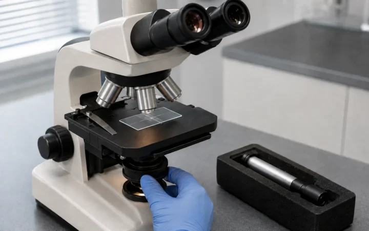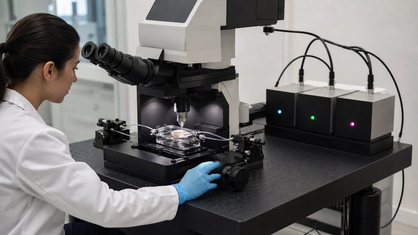

Confocal microscopy is a powerful imaging technique that provides high-resolution, optically sectioned images deep inside biological tissue. In neuroscience, this means visualizing fine neural structures – from single synapses to 3D networks of neurons – with unprecedented claritypmc.ncbi.nlm.nih.govlabcompare.com. Modern confocal systems use focused laser light and tiny pinhole apertures to eliminate out-of-focus blur, enabling 3D reconstruction of brain tissue stacks. As a result, researchers can trace neural circuits in brain slices, observe live calcium signals in neurons, and study glial interactions with sub-micron detail.
2026 shortlist update: Current high-end options include Nikon AX/AX R with NSPARC, ZEISS LSM 910/990, Leica STELLARIS and Evident FLUOVIEW FV5000. For deep in-vivo brain imaging, dedicated multiphoton platforms such as Nikon AX R MP or Evident FV5000MPE belong in a separate decision category. The strongest choice depends on live-cell speed, phototoxicity, spectral flexibility, super-resolution needs, imaging depth, local service and the total configured workflow—not brand name alone.
Table of contents
2026 Confocal Microscope Decision Guide
| System | Strong fit for | Why it stands out in 2026 | Important consideration |
|---|---|---|---|
| Nikon AX / AX R + NSPARC | Fast multicolor confocal imaging, large fields, super-resolution | NSPARC image-scanning microscopy, FN25 field, resonant scanning on AX R and broad visible/NIR laser configuration | Use AX R MP when multiphoton deep-tissue imaging is central to the project |
| ZEISS LSM 990 | Multimodal imaging, fast volumetric live imaging, spectral multiplexing | Airyscan super-resolution, Lightfield 4D and advanced spectral options on the current LSM platform | Choose the configuration around the experiment; not every lab needs every multimodal option |
| Leica STELLARIS | Spectral imaging, fluorescence-lifetime workflows and live-cell microscopy | Power HyD detectors, White Light Laser options, TauSense/FLIM and expandable advanced modalities | Detector, laser and modality choices vary substantially by configuration |
| Evident FLUOVIEW FV5000 | Quantitative confocal imaging and fast dynamic acquisition | SilVIR photon-counting detection with 2K resonant and 8K galvo scanning | FV3000 and FV4000 are previous generations; use FV5000MPE when multiphoton is required |
| Spinning-disk confocal | Long-term live-cell imaging and rapid neuronal dynamics | Parallel acquisition can provide high frame rates with lower light dose than point scanning in many workflows | Optical sectioning and performance depend strongly on the disk, detector and sample |
| Multiphoton platform | Deep brain tissue and in-vivo imaging | Near-infrared nonlinear excitation is better suited to deeper, scattering specimens | It is a different imaging class from conventional confocal and adds workflow complexity |
To choose the best confocal microscope for neuroscience, we examine key imaging needs (speed, resolution, depth, multi-color, live-cell imaging, etc.) and compare current systems from leading manufacturers (ZEISS, Nikon, Leica Microsystems and Evident) alongside spinning-disk and multiphoton alternatives. We highlight each system’s strengths, give real-world examples, and link to expert guides (e.g. on digital imagingfreditech.com and lab equipment selectionfreditech.com).

How Confocal Microscopy Works (Basics)
A confocal microscope scans a focused laser beam across a sample and uses a pinhole aperture to reject out-of-focus light. The key principles:
- Point illumination and detection: The system directs laser light to a small focal spot in the tissue. Emitted fluorescence from that spot passes back through a pinhole aligned to the focal plane. Out-of-focus fluorescence is blocked, drastically improving image contrast.
- Optical sectioning: By scanning point-by-point (or line-by-line) and rejecting blur, confocal microscopes capture “optical slices” at different depths. Stacking these slices builds a 3D reconstruction of the sample.
- High resolution in thick samples: This design allows high-resolution imaging in thick, scattering brain tissue, unlike wide-field fluorescence microscopy. The technique was invented by Marvin Minsky in the 1950s.
- Multiple modalities: Modern systems often add multiple lasers (for multi-color imaging) and detectors (GaAsP photomultiplier tubes or hybrid detectors) for higher sensitivity. They may also include resonant scanners or spinning disks for faster imaging.
Why this matters for neuroscience: Brain tissue is often thick and autofluorescent. Confocal’s optical sectioning lets neuroscientists image dendritic spines, axonal projections, and intracellular calcium signals in three dimensions without hardware sectioning. For example, confocal imaging of cleared mouse brain sections reveals the 3D organization of neuronal layers, and time-lapse confocal movies can track synaptic vesicle dynamics in live neurons.

Figure: A researcher uses a Zeiss confocal microscope (LSM 5 Live) to image live neurons in a culture dish. Confocal optics allow visualization of detailed neural structures deep within the sample.
Key Advantages for Neuroscience
Confocal microscopy offers several advantages that are highly valuable in neuroscience research:
- 3D visualization of neural circuits: By stacking optical sections, confocal microscopes can reconstruct the complex 3D architecture of brain tissue. Researchers can trace neuron morphologies, map synaptic connections, and build detailed brain atlases. For instance, confocal imaging is widely used in “connectomics” studies to map networks in Drosophila or mouse brain slices.
- High contrast and resolution: Laser illumination plus pinhole filtering yields images with high contrast and sub-micron resolution. This makes it ideal for distinguishing fine neural features like dendritic spines, synaptic boutons, or glial processes. A confocal can resolve structures down to ~200 nm laterally (XY) and ~500 nm axially.
- Low background imaging: The pinhole dramatically reduces background blur, ensuring only in-focus fluorophores are imaged. This is crucial in thick brain slices, where autofluorescence and out-of-focus signals would otherwise swamp the image.
- Live-cell compatibility: Confocal systems can image both fixed and live samples. By using sensitive detectors and fast scanning (or spinning-disk scanning), neuroscientists can perform time-lapse imaging of living neurons. Specialized modes (resonant scanners, hybrid confocals, etc.) speed up imaging, enabling real-time observation of calcium waves, protein trafficking, or neuronal firing (via fluorescent indicators).
- Multi-color imaging: Confocals typically have multiple laser lines and filter sets, allowing simultaneous imaging of different fluorophores (e.g. GFP, RFP, calcium dyes). This lets researchers label multiple neuronal populations or molecular markers in the same sample.
- Quantitative analysis: The sharp, well-defined images from confocal microscopes support quantitative measurements (colocalization, fluorescence intensity, volume rendering). For example, one can precisely measure synapse density or neurite branching.
As Dana Smith notes, confocals “collect sharply defined, clear images with low background,” making them excellent for quantitative analysis. They also support both fixed tissue and live imaging, which is essential for neuroscience workflows. In practice, a neuroscientist might use confocal imaging to measure the length of hippocampal dendrites, quantify cell migration in brain organoids, or assess neural stem cell integration in vivo. Such real-world examples abound: confocal studies have illuminated amyloid plaque spread in Alzheimer’s models, neural crest cell migration patterns, and calcium dynamics in retinal cells.
Confocal Microscopy Modes: Choosing the Right Scanning Method
Confocal microscopes come in several flavors, each with trade-offs. The two main types are:
- Laser-Scanning Confocal (LSCM): A focused laser beam (or multiple beams) is scanned across the sample point-by-point (or line-by-line) while detectors capture the emitted fluorescence. This is the classic confocal design. Advantages: Excellent optical sectioning and resolution even in thick samples. Multiple lasers and detector arrays allow flexible multi-color imaging. It is well-suited for 3D reconstructions and high-detail imaging. Downsides: Slower scan speeds; long dwell times per pixel can cause photobleaching/phototoxicity during live imaging.
- Spinning Disk (Nipkow Disc) Confocal: Uses a spinning disk with an array of tiny pinholes to scan multiple points in parallel. This enables very fast imaging with minimal delay. Advantages: High frame rates allow rapid live-cell imaging and virtually no motion blur; lower phototoxicity because each point is illuminated less time. Good for time-lapse of neuronal activity. Downside: Lower optical sectioning for a given pinhole size and reduced depth penetration; typically fewer lasers/modes; needs very sensitive cameras.
- Hybrid or Multi-Mode Confocals: Some systems (e.g. Zeiss Airyscan, Leica Lightning) use a hybrid approach that blends confocal pinholes with detector arrays to boost resolution and speed. These can achieve super-resolution (<150 nm) with built-in deconvolution, at the cost of complexity.
- Multiphoton (Two-Photon) Microscopes: Though technically not a confocal microscope (no pinhole is needed), two-photon systems are often mentioned alongside confocals. They use longer-wavelength IR lasers that excite fluorescence only at the focal point. This allows much deeper tissue penetration (up to ~1 mm in brain) and further reduced photodamage. Multiphoton is often the preferred choice for in vivo imaging through skull or of thick samples, but we discuss it below as an alternative mode in neuroscience.
Choosing between them: Laser-scanning confocals are versatile “all-rounders” good for fixed samples and moderate-speed live imaging. Spinning-disk systems excel in high-speed live imaging (e.g. recording synaptic activity or calcium transients). For deep imaging (brain slices, live animals) or intravital imaging, two-photon may outperform confocal. Hybrid confocals offer a balance: for example, Zeiss’s Airyscan confocal can achieve ~120 nm lateral resolutionpages.zeiss.com. We will mention top products from all categories below.
Laser-Scanning Confocal Advantages
- High resolution & sectioning: Close to diffraction-limited imaging (best-case ~200 nm). You can adjust pinhole size for thinner optical sections, improving axial resolution (though at cost of light).
- Flexible imaging: Compatible with many fluorophores and wavelengths; can image deep via optical sectioning.
- Quantitative clarity: Good for measuring static structures (cell morphology, synaptic density).
- Mature technology: Extensive software, options for automation, FRAP, FRET, etc.
Spinning Disk Confocal Advantages
- Speed: Captures many points in parallel. Entire images are collected in milliseconds. Ideal for capturing fast neuronal events or scanning large areas rapidly.
- Low phototoxicity: Each spot is illuminated briefly, reducing sample damage. Crucial for sensitive neurons.
- Large field of view: Many modern disks (e.g. Yokogawa CSU-W1) offer ultra-wide fields and multiple pinhole sizes for versatilitymicroscope.healthcare.nikon.com.
Multiphoton Imaging (brief note)
- Deep penetration: Using 2-photon or multiphoton can reach hundreds of microns into tissue with minimal out-of-focus damage. Very useful for in vivo brain imaging (e.g. through cranial windows).
- Thicker samples: If you need to image intact brain tissue or live animals, multiphoton is often superior to confocal. Many systems (e.g. Nikon AX R MP) combine confocal and multiphoton modesbiocompare.com.
Choosing the Right Confocal Microscope: Key Features and Considerations
When shopping for a confocal microscope in neuroscience, consider the following factors:
- Imaging Needs: Determine your primary applications. Are you imaging fixed brain slices (where highest resolution matters) or doing live neuronal imaging (speed and low toxicity)? Do you need multi-color capability or fluorescence lifetime imaging (FLIM)? Your answers guide the type of confocal (laser vs disk) and needed options.
- Resolution & Sectioning: Look at the numerical aperture (NA) of the objectives and detector/pinhole capabilities. Some systems (e.g. Zeiss LSM with Airyscan or Leica Lightning) offer super-resolution down to ~120 nm. High-NA water- or oil-immersion objectives can enhance deep imaging and resolution.
- Speed: For live neural activity (fast calcium events, neurophysiology), imaging speed is critical. Spinning-disk units and resonant scanners on laser confocals improve frame rates. Nikon’s resonant scanning or Zeiss’s Airyscan MultiDimension mode can capture hundreds of frames/second.
- Light Sensitivity: Neuroscience often uses weak fluorescent signals (e.g. genetically encoded indicators). Highly sensitive detectors (GaAsP PMTs, hybrid detectors, sCMOS cameras for disk systems) boost image quality with less light.
- Depth Penetration: If imaging deep (thick brain sections, cleared tissue), consider multiphoton capability or infrared lasers. For pure confocal, look for NIR detectors (as in the Nikon AX/AXR series) which help in deep tissue.
- Software & Integration: Good software (ZEN, NIS-Elements, LAS X) is vital for controlling experiments and analyzing data. Also consider compatibility with existing lab software (image analysis pipelines, AI tools). Modular systems with open architecture (e.g. open stages, third-party module compatibility) allow future upgrades.
- Cost & Support: Research confocal systems are usually configured to order. Compare equivalent hardware, software, service, training and upgrade paths rather than relying on generic web price ranges. Local service response and long-term parts/software support can be as important as headline specifications.
For more on selecting lab instruments, see our guide on choosing lab equipmentfreditech.com. When in doubt, prioritize what matters most (e.g. speed vs resolution vs depth) and look at the tradeoffs explained above.
Top Confocal Microscope Systems for Neuroscience
Now let’s review leading confocal microscope models that neuroscientists are using. We focus on systems known for neuroscience or offering cutting-edge features. Included are laser-scanning and spinning-disk systems, with notes on pricing/links where available. (Pricing is approximate and varies by configuration.)
ZEISS LSM 910 / LSM 990 with Airyscan
ZEISS’s current LSM 910 and LSM 990 platforms should lead a 2026 buying discussion rather than the older 900/980 generation. The LSM 990 is the higher-end multimodal platform, combining confocal imaging with Airyscan super-resolution, spectral multiplexing and optional Lightfield 4D. ZEISS currently specifies 90 nm super-resolution for LSM 990. LSM 900 and 980 remain relevant in installed systems and can support newer workflows through compatible upgrades.
- Airyscan: Current LSM 990 configurations support Airyscan super-resolution down to about 90 nm, depending on the optical setup and experiment.
- Fast live-volume imaging: ZEISS Lightfield 4D is available on LSM 910/990 and can reach up to 80 volumes per second in supported configurations.
- Spectral multiplexing: LSM 990 supports advanced spectral separation for complex multicolor experiments.
- ZEN ecosystem: ZEISS integrates acquisition, multimodal workflows and downstream analysis through ZEN, with configuration-dependent tools for live imaging and advanced experiments.
Neuroscience relevance: LSM 910/990 is particularly compelling when a lab needs to combine high-resolution confocal imaging with fast 3D dynamics, spectral separation or multimodal experiments. Lightfield 4D is specifically positioned for fast physiological and neuronal processes while reducing repeated illumination.
- Pricing: Treat ZEISS pricing as quote-based. Final cost depends on stand, detectors, lasers, Airyscan/Lightfield options, objectives, environmental control, software, service and regional support.
Figure: A legacy ZEISS LSM 5 Pascal setup. It remains useful historical context, while current 2026 purchasing decisions should focus on the LSM 910/990 generation and the configuration required by the experiment.
Nikon AX / AX R with NSPARC
Nikon’s AX and AX R are current point-scanning confocal systems, with the optional NSPARC spatial-array detector adding image-scanning-microscopy super-resolution. Nikon positions AX/AX R as its core confocal platform, while AX R MP is the separate multiphoton platform for deep in-vivo and thick-tissue work.

- NSPARC resolution: Nikon specifies approximately 100 nm lateral and 300 nm axial resolution for NSPARC under its stated test conditions.
- Large field and scan range: AX/AX R uses an FN25 scan head and supports large scan formats; AX R adds high-speed resonant scanning for dynamic samples.
- Laser flexibility: Current specifications allow up to 16 visible/NIR wavelengths in supported configurations.
- Confocal vs multiphoton: AX/AX R is the confocal line. For dedicated multiphoton deep imaging, Nikon offers AX R MP with NSPARC.
- AI-assisted workflow: NIS-Elements includes AI-assisted acquisition and processing tools, while the hardware remains modular and upgradeable.
Neuroscience relevance: AX R is a strong fit for fast multicolor neuronal imaging when resonant scanning, a large field of view and low-light super-resolution are priorities. AX R MP should be evaluated separately when the scientific question requires deep multiphoton imaging.
- Pricing: Nikon systems are configured to order. Compare quotes using the same detector count, laser set, objectives, environmental control, software, service term and whether multiphoton hardware is included.
Leica STELLARIS (Current) / SP8 (Legacy)
Leica STELLARIS is the current Leica Microsystems confocal platform; SP8 remains common in established imaging cores but should be treated as the earlier generation in a 2026 buying guide. Leica Microsystems is a Danaher company. STELLARIS emphasizes sensitive spectral detection, flexible laser excitation, lifetime-enabled imaging and modular integration with advanced modalities.
- Power HyD detectors: STELLARIS uses the Power HyD detector family for sensitive spectral detection, including supported far-red/NIR configurations.
- White Light Laser: WLL configurations can tune excitation to the fluorophores used instead of forcing experiments around only fixed laser lines.
- Lifetime and super-resolution: STELLARIS can integrate TauSense/FLIM, LIGHTNING and STED workflows, depending on configuration.
- Live-cell and intelligent acquisition: Current STELLARIS options include AI denoising and autonomous/assisted microscopy tools intended to improve acquisition efficiency and reproducibility.
- Deep imaging options: STELLARIS can be configured with multiphoton-related modalities when deep-tissue imaging is part of the research program.
Neuroscience relevance: STELLARIS is especially useful for labs that need flexible spectral separation, fluorescence-lifetime information, sensitive live-cell acquisition or a path to modalities such as STED and multiphoton imaging. Existing SP8 systems remain scientifically useful, but they should not be presented as Leica’s primary new-purchase platform in 2026.
- Pricing: Leica configurations are quote-based. Detector count, White Light Laser options, lifetime/super-resolution modules, objectives, incubation, software and service can materially change total cost.
Evident FLUOVIEW FV5000 (Replaces FV3000 / FV4000)
The current Evident FLUOVIEW FV5000 replaces the FV4000, FV3000 and earlier FV-series confocal systems. That makes FV5000 the appropriate platform to lead this section in a 2026 buying guide. Older Olympus-branded FV3000 systems remain relevant in installed laboratories, but Evident lists FV3000 as discontinued.
- SilVIR detection: FV5000 uses SilVIR detectors for low-noise, photon-level quantitative acquisition across dim and bright signals.
- Fast and high-resolution scanning: Evident lists 2K resonant and 8K galvo scanning on the FV5000 platform.
- Automation and reproducibility: The current platform emphasizes smart automation and stable quantitative imaging for repeatable experiments.
- Multiphoton path: For deep in-vivo and thick-tissue work, FV5000MPE is the dedicated current multiphoton platform.
- Legacy guidance: FV3000 and FV4000 workflows remain relevant to labs that already own them, but they should not be described as Evident’s current flagship purchase options.
Neuroscience relevance: FV5000 is a current option for labs prioritizing quantitative fluorescence, sensitive detection and fast confocal acquisition. Researchers working mainly on deep living brain tissue should compare the FV5000MPE rather than assuming the standard confocal configuration is the best match.
- Pricing: Evident systems are quote-based; compare detector, laser, objective, automation, environmental-control and service configurations on like-for-like terms.
Spinning Disk Confocals (Nikon CSU-W1, Andor/Yokogawa)
For high-speed live imaging of neurons, spinning-disk systems are top performers:
- Nikon CSU-W1 Yokogawa: This system offers dual-disk (25 μm and 50 μm pinholes) flexibility. Key benefits: ultra-wide field of view (4× larger than older CSU-X1 models) for scanning large brain cultures, and reduced crosstalk for thick samples. It supports two camera ports for simultaneous dual-color imaging.
- Andor Revolution / VisiTech Yokogawa: Andor (now Oxford Instruments) and Yokogawa produce similar spinning disk units (e.g. CSU-X1, CSU-W1). These are often sold with various microscopes or in stand-alone setups.
- Olympus CSU-X1: Another widely used disk unit, often paired with Olympus microscopes for in vivo or fast imaging.
Example: Spinning disk confocals are a go-to for imaging fast calcium waves in cultured neurons or developing zebrafish brains. For instance, the Yokogawa CSU-W1 has been used to image neuronal activity at video rates with minimal photobleaching, enabling hours-long live experiments.
- Pricing: Spinning-disk system cost depends heavily on the microscope stand, disk head, camera or cameras, lasers, environmental control and software. Request a quote that includes the complete live-imaging workflow rather than comparing the disk module alone.
Other Notable Systems
- Bruker Ultima / Prairie Technologies: Known for multiphoton, but also offer confocal options on the same platform.
- Tissue Imaging Platforms: Companies like TissueGnostics (TissueFAXS) and 3i (Intelligent Imaging Innovations) make integrated systems for scanning slides or small animals, blending confocal with automated stages.
- Emerging: Light Sheet Confocal Hybrids: Not strictly confocal, but some new systems (e.g. Nikon LightSheet with confocal capabilities) may emerge as hybrid solutions for brain imaging.
Each lab’s “best” system depends on budget and needs. The major players (Zeiss, Nikon, Leica, Olympus) dominate the market, which means strong support and software. Many core facilities offer training on these instruments – it’s often wise to test your imaging needs on different systems before buying.
Budget and Cost Considerations
Confocal microscopes are major capital purchases, but fixed web price ranges age quickly because research systems are configured to order. A useful budget comparison starts with a like-for-like specification rather than a headline chassis price.
- Hardware configuration: compare microscope stand, scan head, detector channels, laser lines, objectives, motorized stages and environmental control.
- Advanced modalities: Airyscan/NSPARC, FLIM, STED, light-field imaging, spectral modules and multiphoton capability can change project scope substantially.
- Software and computing: acquisition licenses, analysis modules, GPU workstations, storage and data-management requirements belong in the budget.
- Service and uptime: compare warranty length, preventive maintenance, response time, local engineer coverage and post-warranty service.
- Legacy/used systems: a lower acquisition price can be attractive, but confirm detector condition, laser life, software support, parts availability and serviceability before purchase.
For procurement, request comparable quotes from each vendor using the same experimental requirements. Ask vendors to separate essential components from optional upgrades so you can see which features directly support your neuroscience workflow. Also include training, relocation constraints, room requirements and expected data-storage growth.
If the instrument will not be used heavily enough to justify ownership, a microscopy core can be more economical and may provide access to several platforms plus trained staff. Core-facility hourly fees vary by institution, instrument and user type, so compare local access before purchasing a dedicated system.
Conclusion
In 2026, there is no single confocal microscope that is optimal for every neuroscience lab. Nikon AX/AX R with NSPARC is compelling for large-field, fast and super-resolution confocal work; ZEISS LSM 910/990 is strong for multimodal, spectral and fast volumetric workflows; Leica STELLARIS offers flexible spectral, lifetime and advanced-modality integration; and Evident FV5000 brings current-generation quantitative detection and fast scanning. For rapid long-term live imaging, spinning-disk systems remain highly relevant, while deep in-vivo brain imaging often points toward a dedicated multiphoton platform.
The purchase decision should begin with the experiment: required depth, temporal resolution, fluorophore count, phototoxicity tolerance, field of view, quantitative needs and local support. Build a short list from those constraints, test representative samples on candidate systems when possible, and compare quotes using equivalent configurations. For related planning, see our guides to choosing lab equipment and advanced imaging techniques.
FAQ (Frequently Asked Questions)
What is a confocal microscope and how does it differ from a regular fluorescence microscope?
A confocal microscope uses focused laser light and a pinhole aperture to reject out-of-focus fluorescence, enabling sharp optical sections. Unlike a wide-field fluorescence microscope (which illuminates the whole sample at once), confocal scans point-by-point and blocks background light. This yields much higher contrast and 3D imaging capability, which is crucial for thick brain samples.
Why is confocal microscopy important in neuroscience research?
Confocal enables high-resolution 3D imaging of neural structures deep within brain tissue. It can image single synapses or entire neural networks by stacking optical sections. This lets researchers map neural circuits, track neuron development, and quantify changes in disease models. Its ability to image live neurons over time is also invaluable for studying dynamic processes like calcium signaling.
What’s the difference between laser-scanning and spinning-disk confocal?
Laser-scanning confocals use one (or a few) laser spots that raster-scan the sample; they offer excellent resolution and flexibility but are slower. Spinning-disk confocals use many pinholes on a rotating disk to scan in parallel. Spinning disks are much faster (allowing video-rate imaging) and gentler on live cells (less phototoxicity), but usually provide slightly lower optical sectioning. Choose laser-scanning for highest detail, spinning-disk for fast live imaging.
How do I choose a confocal microscope for my lab?
Start with the sample and experiment: tissue thickness, live vs fixed imaging, required speed, fluorophore count, phototoxicity tolerance, field of view and whether super-resolution or multiphoton imaging is essential. Then compare detector sensitivity, objectives, scanner options, software, service and upgrade path. Testing your own representative sample on shortlisted systems is more informative than comparing specification sheets alone.
How much does a confocal microscope cost?
Research confocal systems are usually configured to order, so a single web price is rarely meaningful. Total cost depends on detectors, lasers, objectives, scanner type, environmental control, advanced modalities, software, workstation/storage and service. Ask vendors for comparable itemized quotes and include lifecycle support—not just acquisition price—in the decision.
Can confocal microscopes do live imaging of neurons?
Yes, many confocals are designed for live imaging. Spinning-disk confocals excel at this due to low photodamage. Laser-scanning confocals with resonant scanners can also capture live cell movies. Key is to use low laser power and fast acquisition. Environmental controls (stage top incubator) keep neurons alive. Confocal live imaging has been used to watch neuron firing (via calcium indicators), axonal transport, and synapse formation.
What’s the difference between confocal and two-photon microscopy for brain imaging?
Both provide optical sectioning, but two-photon uses infrared light to excite only the focal plane, allowing deeper tissue imaging (up to ~1 mm in brain) and even less photodamage. If you need to image through scattering tissue or in vivo (e.g., mouse brain under a window), two-photon often outperforms confocal. However, confocals are simpler to use and suffice for cultured neurons or thin slices. Some systems (e.g. Nikon AX R) combine both.
Which brands are best for neuroscience confocals?
Major current research-microscopy manufacturers include ZEISS, Nikon, Leica Microsystems (a Danaher company) and Evident (the current brand behind the FLUOVIEW line formerly associated with Olympus Scientific Solutions). Each platform emphasizes different combinations of speed, spectral flexibility, super-resolution, quantitative detection and deep imaging. The right choice depends on the lab’s experiment design, service access and configured system rather than brand name alone.